Iodination Consultancy Group | Technical information for iodinating proteins and peptides with iodine 125
Technical information on the most common methods for the iodination of proteins, peptides and chemical compounds that are susceptible to iodination with iodine 125. Further information on trouble-shooting procedures for failed iodination reactions and suggestions to increase the yield and therefore efficiency of labelling reactions.
iodine 125, iodination, chloramine t, lactoperoxidase, bolton and hunter, iodogen, purification, sec, reverse phase hplc, ion exchange, chromatography, x-ray, gamma, radiation,
15373
page-template-default,page,page-id-15373,ajax_fade,page_not_loaded,,qode-theme-ver-9.2,wpb-js-composer js-comp-ver-4.11.2.1,vc_responsive
 

Technical

Summary
125I is widely used in the preparation of tracers for sensitive immunoassays and other procedures for the detection, localisation and quantitation of substances in biological samples, for example, receptor binding assays. Radioiodine can be introduced into proteins/peptides either directly- using a soluble oxidising agent such as chloramine-T, or indirectly by conjugating to a ready labelled compound such as 125I-Bolton & Hunter reagent. Indirect or conjugation methods are milder and avoid exposure of proteins to potentially denaturing conditions during the labelling procedure. Following labelling, the tracer should be carefully purified to remove unreacted 125I and other reaction by-products. Before use, the properties of the tracer should be investigated to confirm that it reflects the same biological activity of the unlabelled material.
Tyrosine complex labelled with [125I]Iodine

Factors for consideration
Introduction of a ‘foreign’ iodine molecule into a protein or peptide may alter it’s biological behavior (in contrast to 14C or 3H labelling where the radioactive nuclide usually replaces a naturally occurring non-radioactive isotope of the same element found in the native molecule). The labelling procedure itself may cause considerable changes in protein structure and hence biological activity. However it is possible that one biochemical activity of a protein may be destroyed while a second biochemical activity is unaffected, for example, labelled hormones may lose their ability to bind to receptor sites but not their ability to bind to antibodies. Therefore it is vital to consider the biochemical properties of both the unlabelled and labelled versions of the protein when deciding on its validity during an experiment.

Why choose 125I?
125I is a (low-energy) gamma emitting radionuclide and therefore offers great advantages when it comes to counting samples for radioactive content. No special sample preparation is required which is of particular importance when large numbers of samples are involved. 125I can be easily introduced into the tyrosine residues of polypeptides and this offers the great advantage of a much higher specific activity over the common Beta-emitting isotopes 3H and 14C. For example, one gram atom of 125I gives approximately a hundred times the count rate of one gram atom of 14C. 125I has a half-life of 59.6 days and has a high specific activity at 100% isotopic abundance. 125I can be counted in well-type sodium iodide crystal scintillators with a high counting efficiency (>70%) for accurate quantitation of samples.

Mechanisms of iodination
The most widely used and best understood methods of iodination involve the oxidation of Na 125I in the presence of a protein (or other molecule) containing a tyrosine molecule with the subsequent incorporation of the radioiodine into the tyrosine. Some iodine may also react with histidine, tryptophan or sulphydryl groups, but tyrosine is the principle amino acid involved. Different tyrosine residues present in a protein demonstrate different degrees of reactivity depending on the micro-environment of each individual side-chain in the protein with the residues on the surface of the protein iodinating the most readily. An example of this can be seen in the labelling of insulin where the tyrosine residue in position 14 on the A chain is iodinated preferentially over the tyrosine residue in position 26 on the B chain. By manipulating the reaction conditions, this situation can be reversed.
The chloramine-T method of iodination is probably best understood although other methods of oxidative (direct) labelling along with conjugative methods exist and will be discussed here.

Chloramine-T method
Chloramine-T (a sodium salt of the N-monochloro derivative of p-toluene sulphonamide) breaks down slowly in aqueous solutions forming hypochlorous acid (a mild oxidising agent). In the presence of chloramine-T and under slightly alkaline conditions (pH 7.5), Na 125I is oxidised forming cationic iodine (I+) which is readily taken up by available tyrosine residues in the protein. The iodine atoms substitute ortho to the hydroxyl group on the phenolic ring of tyrosine. Minimal quantities of chloramine-T should be employed to utilise the required amount of isotope as higher concentrations are a potential cause of decomposition of the protein. Reactions for simple proteins are almost instantaneous thus minimising the exposure of the proteins to potential damage. More complex proteins can be labelled using chloramine-T although incorporation of iodide is more time dependent and a given incorporation can be achieved by using lower concentrations of chloramine-T but with longer reaction times. Above pH 8 – 8.5, the substitution of iodine into the imidazole ring of histidine is favoured which can be useful if the tyrosine residues are present within the biologically active portion of the protein. A high incorporation rate is normally obtained although many proteins appear to be readily damaged by this method and enzymatic activity may even be lost after exposure to oxidising agents.

Iodogen (an insoluble oxidising agent) method
Iodogen (1,3,4,6-tetrachloro-3a, 6a-diphenylglycouril) is water insoluble and may be coated onto the surface of a reaction vessel. Iodination is initiated by the addition of Na 125I and buffered protein solution to this vessel. A reaction is allowed to proceed for a predetermined amount of time (usually 5-10 mins) after which time it can be terminated by the addition of excess reducing agent or by removal by aspiration of the reaction mix from the reaction vessel. Disadvantages of this method include longer reaction times and a lower incorporation of iodine into protein molecules. Obvious advantages are that the protein is not exposed to soluble oxidative agents and the reaction mix can be removed from the oxidising agent after the desired reaction time.

Enzymatic iodination
The enzyme lactoperoxidase can be used to catalyse the oxidation of iodide in the presence of very small amounts of hydrogen peroxide. This method is widely used for the preparation of tracers for radioimmunoassays. The protein is mixed with Na125I and lactoperoxidase and the reaction is initiated by the addition of hydrogen peroxide which can be maintained by further additions. The reaction can be terminated either by adding cysteine (providing the protein contains no disulphide bridges) or by dilution. Reactions can be optimised by altering enzyme or hydrogen peroxide mass and by time. Rate of iodination varies greatly at different pH values and the properties and stability of the protein should be taken into consideration when choosing a pH. Enzymatic labelling has shown decreased levels of damage to labelled proteins thus preserving their immunological and other biological activities. Disadvantages include low yields of the iodination reaction thus making it difficult to obtain radioiodinated proteins of high specific radioactivity. In addition, some of the enzyme itself can become radioiodinated and cannot be readily separated from the reaction mixture. The enzymatic method has also been used to label the histidine residues of some proteins.

Conjugation labelling
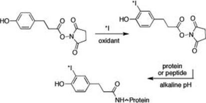
Using N-succinimidyl 3-(4-hydroxy 5[125I] iodophenyl) propionate (Bolton & Hunter reagent) which is commercially available in both the mono-iodinated and the di-iodo form. The N-succinimidyl group of this reagent condenses with free amino groups of peptides (for example: the epsilon amino side-chains of lysine residues) to form a conjugate in which a radioiodinated phenyl group is covalently linked via an amide bond to the peptide. The conjugation reaction takes place under mildly alkaline conditions (pH 8-8.5) and the rate and efficiency of the reaction is highly concentration dependent, so therefore the protein should be dissolved in the minimum volume of buffer that can be handled (as little as 10ul). Typically 5ug of protein in 10ul 0.1M sodium borate buffer pH 8.4 is reacted with 3-5 moles of labelled ester per mole of protein. Reactions are best carried out in an ice bath for 15-30 mins. The reaction mixture can be purified to remove unreacted hydrolysis products and from the low molecular weight iodinated conjugate.
Bolton & Hunter reagent conjugation labelling of free amino groups

This labelling method has the advantage that it overcomes the problems of iodination damage associated with exposure of proteins to oxidising and reducing agents. There is minimal chemical damage to the protein and the labelling site is different from other labelling methods which is useful for proteins that lack suitable tyrosine residues for direct iodination. Using this conjugation method, it is possible to produce labelled enzyme preparations in which the activity is fully retained. It is also possible to produce iodinated tracers with a high retention of immunological activity from unstable proteins highly susceptible to damage from chloramine-T. This is of particular interest when considering iodination methods to be used for proteins that are to be used in Radio Immuno Assays (RIA).

Troubleshooting
Radioiodinated proteins may lose some of their biological activity during the labelling process. This problem may be caused by the substitution of iodine into the tyrosine residues of the protein, or the reagents used in the iodination may modify other labile amino acid residues associated with the biologically active site. If this is the case, it will be necessary to label amino acid residues other than tyrosine or to use a more gentle chemical labelling technique.
In general, iodination reactions are carried out on the microscale with volumes totalling only a few tens to hundreds of microlitres. Scrupulous care and attention to minute detail is required. This detail is required to ensure that the small volumes are handled safely and are mixed adequately which should result in successful iodination and safe working practices. A commonly occurring problem is a reaction where little or no incorporation of iodide has taken place. In the case of no incorporation, the most likely explanation is the omission of either the protein or the oxidising agent or that the reaction  has not been mixed adequately during the iodination. Incorrect pH or sufficient volume of buffer may offer another explanation for little or no incorporation of iodide into proteins. It is also possible that the radioiodide solution may be ageing or substandard in which case, a new batch should be tried. Another cause of a failed reaction may be the quality of the protein itself. Because such minute quantities of proteins are utilised during the reaction, the remaining protein solution  may have to be stored frozen for long periods of time and without any stabiliser or carrier protein. Stocks of proteins for labelling should be stored in small aliquots and should only be subjected to one freeze-thaw cycle, however, we should remember that some proteins are unsuitable to be frozen anyway. Particularly unstable proteins should be stored at -70oC or below. All of the above are only the most common reasons for a failed iodination reaction, there are substantially more causes that can be attributed to failed reactions which are too lengthy to be listed here.

 Iodination yield

None

Immunological activity

Some possible causes

No protein added
No oxidising agent added
Reagent unmixed

Low

Normal

Insufficient oxidising agent
Reaction not buffered to optimum pH
Inadequate mixing of reagents

Normal or impaired

Old or defective radioiodide preparation
Ageing protein preparation

Impaired

Protein not adequately separated from salt after labelling
Non-specific adsorption of labelled protein to reaction vessel

Normal or high

Impaired

Excessive oxidising agent added
Excessive reducing agent added
Old or defective radioiodide preparation
Ageing protein preparation
Excessive incorporation of radioiodide into protein
 Protein not adequately separated from salt after labelling
Reducing agent omitted with consequent iodination of carrier proteins and contamination of tracer

Purification of radioiodination reactions
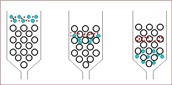
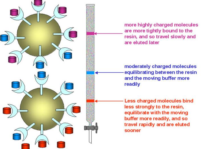
All radioiodination reactions contain undesired by-products (e.g. unreacted iodide, oxidising and reducing agents) that require removal before the labelled protein can be used in an application. Gel-filtration chromatography (Fig.1) is the simplest and most common method used for purification. However, the tracer recovered using gel-filtration chromatography will be contaminated with damaged protein (i.e. multi-iodinated protein, aggregates and oxidised protein) thus requiring additional purification. Methods used for additional purification will depend upon the individual cases. The most powerful separation techniques include affinity (Fig.2) chromataography, ion-exchange and reversed phase HPLC (RP-HPLC) (Fig.3).
Fig.1: Gel filtration / Size-exclusion chromatography (SEC)
Fig.2: Affinity chromatography
Fig.3: Reversed-Phase HPLC (RP-HPLC)

Affinity chromatography is particularly useful when labelling immunologically active materials and receptor ligands. The conditions required to remove labelled the ligand from the affinity material are usually fairly harsh and can damage the radioiodinated protein. Affinity chromatography does not remove or separate the unlabelled material. Ion-exchange chromatography which separates according to charge can be used to separate di-iodinated products and unlabelled material as well as lactoperoxidase but will not separate various multi-iodinated species or oxidised material. Reversed-phase HPLC can be used to separate various mono-iodinated materials from all the other reaction products and yield a pure product at maximum specific activity. The latter technique is ideally suited to small peptides (<8,000 mol.wt).

Determination of Specific Activity
Specific activity (SA) is defined as the unit of radioactivity per mole of protein. In the case of iodine, this is quoted as TBq/mmol or Ci/mmol. Introduction of a single atom of 125I into a protein causes the least alteration to it’s structure and thus keeps to a minimum any substitution effects. In general, further incorporation accelerates radiolytic decomposition . For receptor studies, specific activity must be known accurately as it is used to quantify the density of a ligand and it’s affinity for receptors. In order to calculate the specific activity, the amounts of protein and radioactive iodide used in the reaction need to be known, as well as the yield of the product.
There are several ways to determine specific activity. If gel-filtration is used as a method of purification, unreacted 125Iodide and labelled protein can both be measured in a calibrated instrument. Measurement of radioactivity originally taken and that of the salt peak (unreacted free iodide) allows the calculation of the amount of radioactivity in the protein.
Ascending thin-layer chromatography (TLC) can also be used to determine specific activity using a suitable medium and solvent (e.g. methanol : acetone, 50:50). In using a suitable elution solvent, the labelled protein remains at the origin and the 125Iodide migrates up the plate at the solvent front. By determining the percentage incorporation (the ratio of radioactivity between the two spots on the eluted TLC plate) and knowing the amount of radioactivity and protein used, the specific activity can be calculated.
Trichloroacetic acid (TCA) precipitation can also be used to determine specific activity. A small sample of the reaction is taken, diluted and precipitated in 10% TCA. After 30 minutes on ice, the precipitate is pelleted by centrifugation for 5 minutes at 10,000g. An aliquot of the supernatant is counted and compared with the counts taken before precipitation to determine the level of incorporation in the reaction.

Final formulation and stability of the radioiodinated protein
There are various factors that limit the useful life of a radioiodinated protein, some of which are:
– Radiation (radiolytic) decomposition
– Loss of radioiodide
– Aggregation of labelled protein
– General loss of immunoreactivity of the labelled preparation
In general, commercial radioiodinated proteins are supplied lyophilised from a pH buffered solution in the presence of various bulking agents: (e.g. lactose), a carrier protein (e.g. Bovine Serum Albumen- BSA) and a protease inhibitor (e.g. aprotinin). In some cases where the stability of the radioiodinated compound has been evaluated, the formulations are less costly, i.e. a buffered liquid solution stored at either 2-8oC or -20oC.

Radiation decomposition
The greater the number of radioactive atoms incorporated per molecule of protein, the greater the number of radioactivity decay fragments which could show reduced immunoreactivity. This effect is exacerbated if individual tyrosine residues are di-iodinated. The stability of a labelled protein can be extended by limiting the level of incorporation of radioiodine to one atom per molecule of protein.